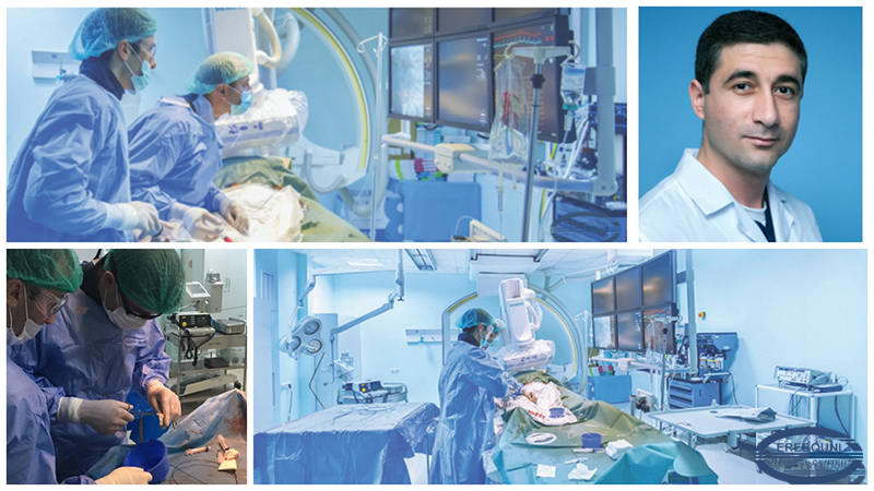
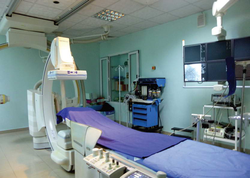
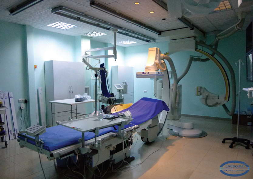
For the first time In Armenia in MC “Erebouni” was performed embolization of giant liver hemangioma by means of endovascular surgery, which is unique in its kind, both in terms of minimal invasiveness of the procedure and complexity , so in its effectiveness.
Cavernous hemangioma is the most common type of benign liver tumor, which is clinically associated with abdominal pain, pains in the right upper quadrant, dyspepsia, constipations and cuagolopathy. Massive hemangiomas are dangerous for the risk of spontaneous rupture and can lead to death. Open surgical intervention is used with frequency of 30% and only in case of the lesion located within one lobe of the liver. Alternative treatment, which was recently implemented to routine clinical practice, is endovascular transarterial embolization of hemangiomas that is effective, gentle, less invasive radical surgery.
A 50-yr-old patient A.G. was admitted to MC “Erebouni” with complaints of pains in epigastric region, in the left upper quadrant of abdomen, dyspepsia, constipations and general weakness in 19.03.16. The patient has suffered from above mentioned complaints for 7 years. Primary it was diagnosed as liver tumor, according to that the patient was supervised by an oncologist. On admission the CT scan was carried out, on base of which it was diagnosed: hepatic hemangiomatosis, massive hemangioma in the left lobe of the liver, the displacement of the organs (as a result of squeezing them by the giant hemangioma). The biggest lesion – 21,2× 17.1× 9,7cm covered the left lobe of the liver, when as lesions of 5,5 × 5,0cm,1,5 × 1,4cm, 3,1 × 2,5 cm and 3,5 × 3,4 cm were situated in the right lobe of the liver.Taking into consideration the distribution of the process it was decided to perform transarterial embolisation of hemangioma.
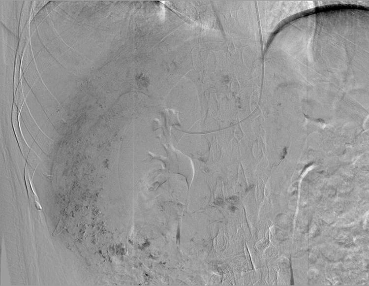
After carrying out clinical and laboratory examination in 19.03.16., the surgery was performed under the local anesthesia by the Head of Angiography and Intervention Cardiology Department Dr. A. Tzaturyan: through the right femoral arterial access was carried out selective digital subtraction angiography (DSA) of celiac trunk and hepatic arteries with special catheter. It was revealed, that the arteries of hemangioma are generated from distal branches of the right and left hepatic arteries.
In parenchymatous phase were distinctly seen vascular beds, which is specific for cavernous hemangioma. After careful angiographic study, selective embolisation of all feeding branches of hemangiomas with particles of cyanoacrylate (300-500 mcm) was carried out. After the procedure of embolisation , the control angiography of artery has shown a complete occlusion and lack of hemangioma.
On the following day the patient was suffered from severe pains in the right hypochondrium, nausea and vomiting. Taking into consideration clinical and instrumental observations, the patient was diagnosed as acute ischemic cholecystitis, which is one of infrequently encountered complications of embolisation of hepatic arteries as a result of spreading of embolisation particles into cystic artery, which require the temporary draining of the gallbladder.
In 21.03.16 percatuneous liver drainage of the gallbladder was carried out, under local anesthesia and US scan by interventional radiologist Dr. A.Galumyan. After the procedure the symptoms have disappeared.
The patient was discharged on the 10th day with in satisfactory condition. At follow-up ultrasound examinations there wasn`t observed blood flow in hemangiomas .
This kind of surgery was performed in Armenia for the first time in MC “Erebouni” and is unique in its kind, both in terms of minimal invasiveness and complexity , so in its effectiveness.


