During August 2016 in Erebouni MC by invasive radiologist of Department of Angiography Dr A. Tsaturyan were performed several endovascular interventions, which have no analogs in Armenia.
- Uterine artery embolisation
- Preoperative embolisation of hemangioma and metastatic melanoma of femoral lymph nodes
- Embolisation of a giant hemangioma
Patient S.E. 1968y/b., has admitted to Erebouni MC with complaints of back pain, frequent urination, uterine bleeding, bloating. Diagnosis: multisite uterine fibroids, uterine bleeding. Uterine sizes are consistent with 20 weeks of pregnancy.
After clinical and laboratory tests, under local anesthesia was performed uterine artery embolisation: the puncture of femoral artery was made, then through it was guided a special catheter, designed for catheterization of uterine arteries. Under angiographic control, the catheter was first introduced into the left uterine artery. After angiography test the embolisation was carried out until “sludge- phenomena”. The right uterine artery was also embolized. After the procedure of embolisation the catheters was removed and the pressure applied on the femoral artery for a few hours. The surgery takes about 30 to 40 min.
Postoperative period was uneventful. The patient stays under supervision of the doctors in hospital. After the procedure of endovascular embolisation of uterine arteries, uterine bleeding wasn`t observed.
Patient G.R., 1990 y/b., has admitted to Erebouni MC for preoperative embolisation of arteries of hemangioma, which occupied the entire right posterior surface of the chest with the transition into pleural cavity, retroperitoneal space and pelvis.
Several years ago the patient had surgery for hemangiomas removal, but the during surgery bleeding starts, and therefore the surgery was stoped. Hemangiomas are charecterized by severe bleedings in open surgeries. The patient was scheduled for open surgery after embolisation of the arteries of hemangioma.
In 11.08.16. patient G.R. was carried out a selective angiography: it was found that hemangioma is fed from the right intercostal arteries with a diameter of 3-4 mm, and from the branches of the right subclavian artery. Selective catheterization of the selected branches and coil embolization was implemented.
In repeated angiographic contrasting the hemangioma wasn`t observed. The patient was discharged on the next day after procedure and was preparing to open surgery for removal of hemangiomas.
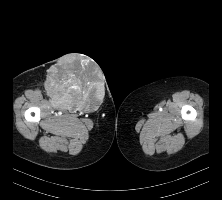
Patient D.A., 1936y/b., in 16.08.16. has admitted to Erebouni MC with diagnosis of metastatic melanoma in the right femoral lymph node, external bleeding from metastasis surface. The patient was planned for surgical removal of metastasis, but because it has a great feeding arterial network, it was decided to carry out preoperative arterial embolisation of metastasis to reduce the risk of bleeding during the surgery.
On the same day, under the local anesthesia, selective arterial angiography of the right lower extremity was performed, whereupon it was found, that metastases have several pedicles, which extend from the middle part of the femoral arteries surface and branches of the deep femoral artery. Under angiographic control it was performed the embolisation of 7 pedicles of metastases, then on control contrast angiography the metastases wasn`t observed. The patient was discharged on the second day and was preparing to open surgery for removal of metastases.
Patient A.M., 1961y/b. has admitted to Erebouni MC in 15.08.16. with complaints of epigastric and the right upper quadrant pain. In anamnesis she has a giant hemangioma of the liver measuring 14×10,5×10,5 cm. This formation was taking the entire left lobe of the liver, partly the right - IV, VII and VIII segments. After clinical and laboratory tests, on the same day the patient was carried out selective angiography of the celiac trunk, in which were detected all pedicles of the hemangioma (the left hepatic artery, several segmental arteries of the right lobe - IV, VII and VIII). All described arteries were embolized with spheres of 300-500 micron.
In repeated contrast angiography no hemangioma has been identified after the procedure. The patient was discharged on the third day in satisfactory condition.
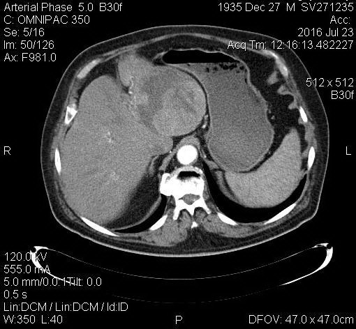
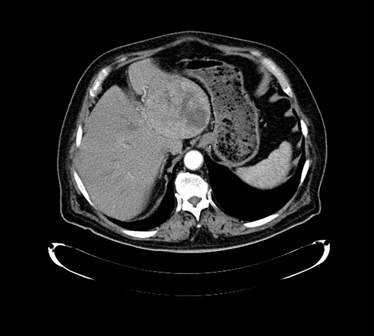
In June 2016 the patient S.V., 1935y/b., has been carried out hepatic arterial chemoembolization because of hepatocellular carcinoma of the left lobe of the liver. The tumor size is 9 × 10 cm. A month later CT –examination was carried out: partial tumor necrosis was observed, without increasing the size and metastatic lesions. To achieve the stabilization of the process, in 18.08.2016 the patient was again carried out the hepatic arterial chemoembolization (Fig. 2 and 3).
Chemoembolization of the hepatic artery is one of the effective methods of regional chemotherapy, performed under local anesthesia, allowing to introduce the chemotherapy medicine directly deep into the tumor, creating it high concentration inside of the tumor tissue, without causing the systemic side effects. Wherein bloodstream within tumor is suspended.
The above interventions are unique and are carried out only in Erebouni MC. By its level of performance they are low-traumatic and minimal invasive, and also are high effective. Intraoperative risk, pain syndrome, recovery time compared to traditional surgery is significantly reduced.


