
A 71-years-old patient L. Sh. , in 30.11.2016 has been admitted to a neck vessel and cardiac surgery department (head - M.P. Shatakhyan, MD, PhD) in a critical condition: the patient didn`t take food during a week because of vomiting when try to swallow, was dehydrated (she was on parenteral nutrition). Because of the extreme weakness she wasn`t able to walk, but movements in the extremities weren`t violated. Earlier the patient complained on pronounced dizziness, uncoordinated movements, headache and nausea.
During physical examination a round solid tumor on the neck, above the left collarbone was allocated. Horner syndrome with a drooping eyelid was also observed. By immediate CT angiography and duplex scanning a large size tumor (5×6 mm) was identified. It was compressing and deforming the left vertebral artery with pronounced blood flow violation. The right vertebral artery was hypoplastic with a diameter of 1.5mm. So, the left vertebral artery was dominant (2.5-3mm) but severally compressed. As a result of compression of the left dominant artery the significant brain ischemia was observed.
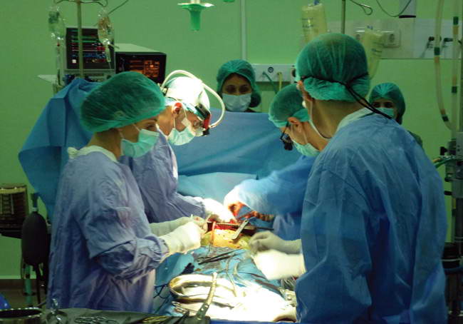
After 24-hour intravenous liquids injection and balance restoration it was decided to perform the surgical intervention to restore patency of the dominant left vertebral artery and provide a complete blood supply to posterior structures of the brain. In 02.12.2016 the surgical team (head of theneck vessel and cardiac surgery department - M.P.Shatakhyan, MD, PhD; surgeons – S.I. Arzumanyan, N.S. Safyan, anesthesiologist - A.R. Muradyan) had perform tumor removal, that was compressing left vertebral artery and sympathetic trunk.
.
Then the vertebral artery was cut off from subclavian artery and relocated anteriorly to the sympathetic trunk, thenanastamosis “end to side” with carotid artery was applied to provide a full blood flow to the brainstem.
Post-operative period was uneventful, without complications. The patient was extubated an hour after the surgery, started eating after 3 hours, walking after 12 hours. After the control duplex scanning of the vessels, which confirmed normal blood flow in the left vertebral artery, the patient was discharged on the 3rd day after the surgery. At follow-up examination on 20 days after the surgery the patient could move independently, eat normally. Dizziness and upper eyelid drooping have disappeared.
The histological analysis of the tumor had revealed a calcified at the edges cavity with long-standing hemorrhagic content. According to the world literature, tumors, compressing the vertebral artery are rare and require the surgical intervention. Such patients usually come to the doctor in serious clinical condition. If the size of the tumor is large, the patients can hardly or even can`t take a food, often suffer from a cerebellar stroke, have a significant dizziness and uncoordinated movements, unable to move. Severe clinical appearance can often give an impression of hopelessness of the surgical intervention. However, after the removal of a mechanical obstruction and restoration of a normal blood flow to a brainstem, the patient is recovering very quickly with restoration of all functions.
