

On March 3, 2017, at a regular Conference of Cardiology Clinic of MC Erebouni was presented clinical case of the patient with Takotsubo cardiomiopathy. Cardiologist of Urgent Cardiology Department Dr. Flora Muradyan made a presentation.
Takotsubo cardiomyopathy was first reported in Japan in 1990 by cardiological community, and already at the European Cardiology Congress 2006 in Barcelona was presented as an independent nosology - as a variation of cardiomyopathy. Takotsubo in translation from Japanese means “the octopus trap”. With this pathology the heart muscle expands and the heart in form becomes more like a vessel (vessel for wine).
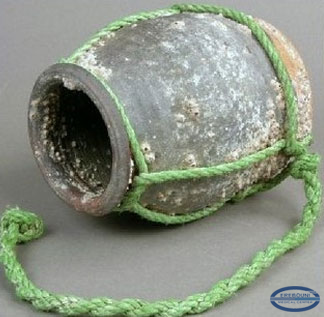
Fig. 1 “ Trap” for octopus
Pathology occurs mainly in middle-aged or older women after severe stress. Hence the names: Transient apical ballooning, Stress-induced cardiomyopathy, Stress cardiomyopathy, “ampulla” cardiomyopathy, Neurogenic myocardial stunning, “Broken heart” syndrome.
The basis of pathogenesis is an increased concentration of catecholamines in blood several times more, than during acute myocardial infarction, which leads to a coronary artery spasm, microcirculatory disorders and death of cardiomyocytes (Y. J. Akashi 2003, Wittstein 2005). Biopsy reveals the signs of intoxication by catecholamines: intracellular accumulation of glycogens, an increase of vacuoles, an increase in the number of proteins in territorial matrix in the absence of myocarditis.
Epidemiology: most often occurs in women >55 (70-90%), during menopause the risk of disease increases almost 5 times. It is also possible the manifestation of the disease in teenagers aged between 14-16, because their body is not very adapted to stress. The overall percentage of this pathology from all cardiac events is 2 %.
The clinical symptoms: In 60% cases is observed acute pain in retrosternal area, short of breathness (in 30 % cases), loss of consciousness, various disorders of heart rhythm, cardiac arrest, development of acute stagnant blood circulation insufficiency (systolic dysfunction, mitral regurgitation III-IV, up to cardiogenic shock), development of left ventricular thrombus and thromboembolic complications.
The diagnosis is based on clinical symptoms, ECG, echocardiogram, concentration of biomarkers and catecholamines, as well as CAG (coronary angiography).
ECG- changes (fig.2,3):
• Elevation of ST-segment in chest leads.
• Prolongation of Q-T interval with inversion of T-wave
• The early recovery of ECG- changes within 2-3 weeks

Fig.2. ECG and ventriculography in Takotsubo cardiomiopathy

Fig.3. ECG differential diagnostics between Takotsubo cardiomyopathy and acute myocardial infarction of anterior localization.
BIOMARKERS AND CATECHOLAMINES. The level of biomarkers of high cardiac sensitivity is much lower, than in myocardial infarction of this volume. The concentration of catecholamines in blood is much higher, than in acute myocardial infarction, however, it should be excluded pheochromocytoma and myocarditis. Echocardiography reveals akinesia or dyskinesia of apical and/or medial segments of LV, hyperkinesis of basal segments of LV, therefore it is also possible obstruction of outflow tract of LV, segmental or total dilation of LV, reduction of ejection fraction of LV from 50% within 4-8 weeks. (Fig.4).

Fig.4. Two-dimensional echocardiography - image Takotsubo cardiomyopathy.
Coronaroangiography reveals the absence of hemodynamically significant coronary arteries constrictions, ventriculography reveals polymorphism of the LV walls movement in the phases of filling and emptying (Fig.5)

Fig.5. Ventriculography with Takotsubo cardiomyopathy.
Treatment. There are no certain guidelines in the treatment of Takotsubo cardiomyopathy. In the acute period the treatment is similar to the treatment of the acute myocardial infarction- analgesics, tranquilizers, antiplatelet agents, ACEI, beta-blockers, diuretics, inotropic drugs, but in case of obstruction of outflow tract of LV inotropic drugs are contraindicated.
Clinical case
In 14.11.2016 in MC Erebouni has admitted a patient S.A., 1956 y/b., with complaints of pain in the chest area, shortness of breath.
Anamnesis: According to ECG - ST-segment elevation in V1-V5 leads. Echocardiography (from 14.11.16.) - LV myocardial hypertrophy, dilatation of the left chambers: LA 4,2cm, LV 5,7 cm. The aorta is indurated, in ascending section is dilated- 4,1cm. Akinesis and hypokinesis of all anteroseptal segments of LV. Myocardial contractility is reduced. Ejection fraction of LV – 27-28%, Doppler: mitral regurgitation of III degree, aortic regurgitation of II degree, pulmonary hypertension II degree, systolic pressure in pulmonary artery Pmax RA 77 mm Hg.
On the base of examinations it was diagnosed: IHD: Acute myocardial infarction of the anterior wall of the LV with ST segment elevation.
In the hospital the patient was detected the development of acute insufficiency of the LV, which was compensated by medicine. The treatment was carried out as in acute myocardial infarction, and was discharged with improvement. After stabilization of health, the patient was recommended coronary angiography, but she had refused. However in 30.11.2016 the patient again appealed to MC Erebouni to carry out angiography - there weren`t revealed any atherosclerotic changes from coronary arteries.
After repeated echocardiogram (16.03.2017.) it was revealed: Myocardial hypertrophy of the LV, dilatation of the left chambers: LA 4,2cm, LV 5,6 cm. The aorta is indurated, in ascending part is dilated - 3,9 cm. Myocardial contractility is reduced. EF-42%. Contractility of the right ventricle is preserved. TAPSE>1,8 cm. Inferior vena cava collapses.
Doppler: mitral regurgitation of II degree, aortic regurgitation of II degree, regurgitation of pulmonary artery II degree, there wasn`t detected pulmonary hypertension. In comparison with previous echocardiograms EF of LV has grown inadequately, because the patient had valvular insufficiency.
Based on clinical and laboratory and instrumental examinations, it was diagnosed: Takotsubo cardiomyopathy, mitral regurgitation of II degree, aortic regurgitation of II degree, valvular insufficiency of pulmonary artery II degree, arterial hypertension, HRreEF, CI FC II ( NYHA):
It is important to note, that for an establishment of more accurate diagnosis of Takotsubo cardiomyopathy is required a certain period of time, which makes it difficult to provide a timely diagnosis of this pathology, since the acute period is progressing as an acute myocardial infarction, and the recovery takes place within a short time (based on ECG and echocardiogram data).
 |
 |
 |
